Fragility Fracture Risk, Common Sites and Treatment Options

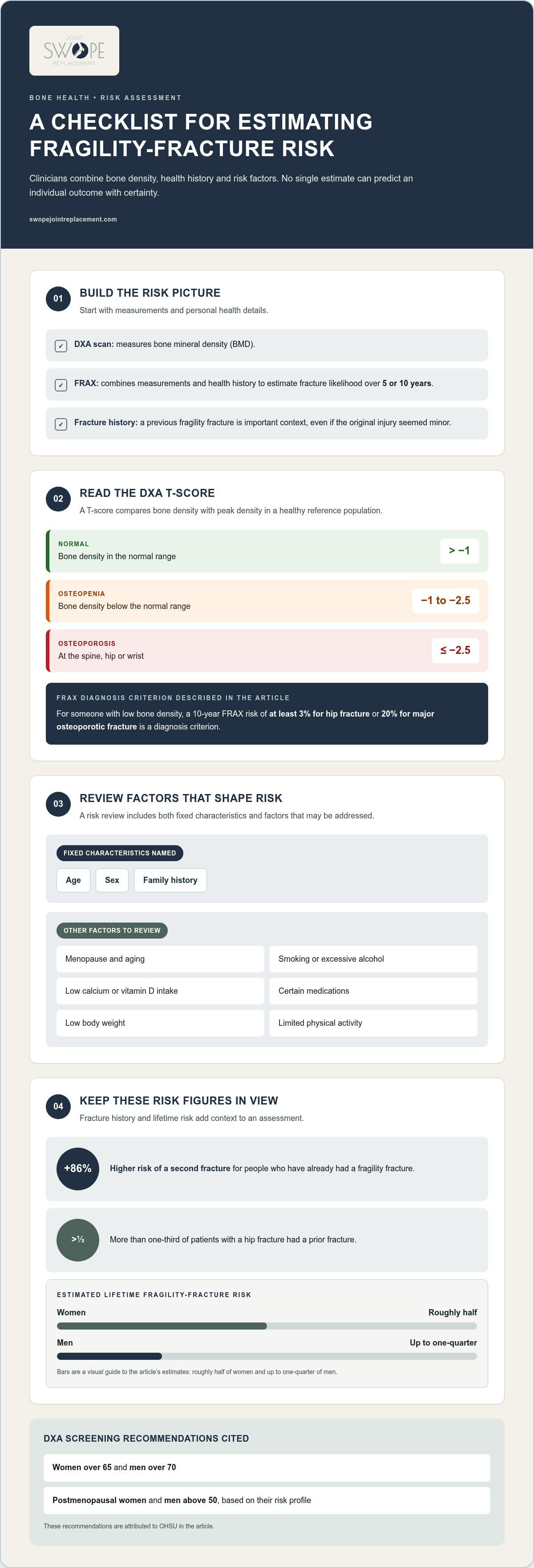
A person’s risk of a fragility fracture can be estimated with tools such as FRAX, which consider health history and measurements to assess fracture risk over five or ten years. A previous fragility fracture matters: people who have had one face an 86% higher risk of a second. These estimates help frame a closer look at bone health and personal risk factors.
It’s understandable to associate fractures with a major fall or accident. Yet a fragility fracture can occur after a fall from standing height or less, or while lifting a light object. The force involved and the condition of the bone help distinguish this injury from other fractures and guide care.
This article explains how clinicians assess fragility fracture risk, where these breaks commonly occur, and how they differ from traumatic and stress fractures. You’ll also learn about treatment options, from immobilization and rehabilitation to surgery, and how addressing bone fragility can help reduce the risk of another break.
Table of Contents
What are the most common sites for a fragility fracture?
A fragility fracture occurs when weakened bone breaks under force that would not usually cause a fracture. It can follow a fall from standing height or less, or lifting a light weight of 5 to 10 pounds. Osteoporosis is the most common underlying cause, though bone cysts, cancer, inherited bone disorders, and osteomyelitis can also weaken bone.
Seek emergency care for a suspected fracture accompanied by visible deformity, numbness, tingling, or marked loss of movement. These symptoms can occur with a break and need prompt assessment, particularly after a fall or while standing.
The hip, spine, and wrist are common locations. Hip fractures can affect standing, walking, and daily activities. They account for about 30% of all osteoporosis fractures. More than 350,000 occur annually in the United States, and from age 85, hip fractures become the most common fracture for both men and women. Assessment considers the injury site and its effect on movement.
Wrist fractures often happen when someone extends a hand to break a fall. More than 300,000 wrist fragility fractures occur every year in the United States. Spinal fractures, also called vertebral fractures, may cause back pain, but symptoms can be subtle. Estimates suggest that as many as three in four vertebral fractures go undiscovered, so a lack of dramatic symptoms does not rule one out.
Osteoporosis-related fractures are associated with low bone mineral density, and their incidence increases after age 50. Imaging used to diagnose a suspected break may include X-rays, MRI, or bone scans. The injury mechanism, symptoms, and affected site help shape assessment, including when back pain raises concern about a vertebral injury.
Hip: A break can limit standing and walking, making mobility central to assessment.
Wrist: A fall onto an outstretched hand is one example of a low-impact event that can injure weakened bone.
Spine: Vertebral fractures may cause back pain, while some produce few noticeable symptoms.
Osteoporosis causes 2 million fractures in the United States each year. Recognizing a fragility fracture at the hip, wrist, or spine helps connect the immediate injury with possible underlying bone weakness. That distinction can guide both fracture care and attention to bone health.
Can an individual’s risk of fragility fracture be predicted?
Risk estimates combine bone density measurements, health history, and other risk factors. Tools such as FRAX help estimate the likelihood of a fragility fracture over five or ten years, but they cannot predict an individual outcome with certainty.
A fragility fracture may follow changes in bone strength associated with age, sex, family history, or menopause. Low calcium or vitamin D intake, smoking, excessive alcohol, and certain medications can also contribute. Previous fractures, low body weight, and a sedentary lifestyle are additional risk factors. Some factors can change, while age and family history cannot.
Diagnosis may involve imaging, such as X-rays, MRI, or bone scans, to identify a fracture. A DXA scan measures bone mineral density, while FRAX combines measurements and health history to estimate future fracture risk.
Factors That Shape Fracture Risk
Risk factors include fixed characteristics, such as age, sex, and family history, as well as factors that can change. Bone disease can develop with aging, and risk is especially relevant for postmenopausal women. Smoking, excessive alcohol, low calcium or vitamin D intake, some medications, low body weight, and limited physical activity can also affect bone health. Reviewing these factors helps identify where prevention efforts may matter.
Assessing and Predicting Fracture Risk
Assessment combines bone density testing with health details to estimate the likelihood of a fracture over five or ten years. A DXA scan measures bone mineral density, and FRAX uses measurements and health history to calculate risk. OHSU recommends DXA exams for women over 65 and men over 70, as well as for postmenopausal women and men above 50 based on their risk profile.
A DXA T-score compares bone density with peak density in a healthy reference population. A T-score greater than -1 is described as normal, while a score between -1 and -2.5 indicates osteopenia. A score of -2.5 or lower at the spine, hip, or wrist indicates osteoporosis. For someone with low bone density, a FRAX 10-year risk of at least 3% for hip fracture or 20% for major osteoporotic fracture is a diagnosis criterion.
Lifetime and Subsequent Fracture Risk
Lifetime estimates show why assessment matters across adulthood. Roughly half of women and up to one-quarter of men will experience a fragility fracture during their lifetime. A previous break also matters: people who sustain one have an 86% higher risk of a second fracture. More than one-third of patients with a hip fracture had a prior fracture, making fracture history valuable in assessment.
Risk estimates can inform decisions, but they are not a diagnosis. Bone density results, previous fractures, health history, and modifiable factors all contribute. A prior break deserves attention even if the original injury seemed minor, because it can signal underlying bone weakness and a higher chance of another fragility fracture.
When a fracture needs orthopaedic evaluation or treatment, trauma and fracture care can address the injury and its effect on movement.
Prevalence of osteoporosis and low bone density in the United States
Osteoporosis weakens bone and increases the chance of a break after modest force. In the United States, bone density measurements help distinguish osteoporosis from low bone mass and guide decisions about bone health.
Bone Density and Bone Strength
Bone mineral density measures how much mineral is present in a specific area of bone, while bone strength reflects its ability to withstand force. A DEXA scan measures density and gives a T-score for comparison with a healthy reference population. A T-score compares a person’s bone density with a healthy reference population. These results help explain why a low-impact event can injure bone.
An estimated 10 million Americans have osteoporosis, and 43.1 million have low bone density. These figures describe different stages of reduced bone strength, not identical diagnoses. Low bone density can signal elevated risk without meeting the threshold for osteoporosis. Considering the result alongside fracture history and health factors gives a more useful assessment than treating one measurement as the whole picture.
Osteoporosis-related fracture sites are associated with low bone mineral density, and fracture incidence increases after age 50. Osteoporosis causes 2 million fractures in the United States each year. A density result can therefore help clarify an injury’s underlying context, especially when a fracture followed a modest impact. It also provides a starting point for discussing whether treatment should address bone strength as well as the break.
Bone Fragility and Osteoporosis Diagnosis
Osteoporosis is diagnosed using bone density criteria, while other conditions can also make bone fragile. A T-score greater than -1 is described as normal, and the range between -1 and -2.5 indicates osteopenia. A T-score of -2.5 or lower at the spine, hip, or wrist meets the osteoporosis threshold. This distinction helps make assessment and treatment discussions more specific.
Osteoporosis is the most common cause of fragility fractures, but it is not the only one. Bone cysts, cancer, inherited bone disorders, and osteomyelitis can also weaken bone. These conditions affect bone in different ways, so identifying the cause matters when planning care. A fracture and a low bone density result may prompt clinicians to consider osteoporosis and other contributing factors.
Bone strength depends on more than a scan result. Calcium, vitamin D, and dietary protein are important nutrients for bone health. Regular exercise and physical activity are among the strategies used to help prevent fractures. These measures support a broader plan, while osteoporosis treatment may address diagnosed bone loss and future fracture risk.
What are the treatment options for fragility fractures?
Care may include immobilization, pain relief, surgery, rehabilitation, and treatment for weakened bone. Complications can include pain, reduced mobility, and difficulty with daily activities. Hip fractures also carry serious health risks.
Medical Treatment and Fracture Prevention
Treatment depends on the fracture’s location, stability, and effect on movement. A cast or brace can immobilize some breaks as they heal, and pain may be managed with over-the-counter or prescription medication. Surgery may be needed to repair or stabilize a fracture, particularly at the hip or spine. Some hip fractures require joint replacement.
Care also addresses bone fragility to help reduce the chance of future breaks. When osteoporosis contributes to a fracture, medication may be prescribed to improve bone density. Options can include bisphosphonates, calcium and vitamin D supplementation, and nutritional support. These measures complement treatment of the injured bone rather than replacing it.
Rehabilitation and Bone Health
Rehabilitation helps a patient regain mobility and function after injury. Physical therapy can support movement and help prevent future fractures, while timely rehabilitation addresses loss of function during recovery. Only 40% to 60% of people who experience a hip fracture recover their prefracture mobility and ability to perform daily activities, according to the U.S. Preventive Services Task Force.
Recovery also involves bone health. Calcium, vitamin D, and dietary protein support bone health and strength. Regular exercise helps maintain physical capacity, while a healthy diet supports the body during healing. The appropriate rehabilitation plan reflects the fracture, the patient’s mobility, and daily activities.
Fall Prevention and Complications
A fall from standing can cause a fracture when bones are weakened, so preventing another fall is an important part of care. Physical activity, strength-building exercise, and attention to nutrition support prevention. OHSU recommends at least 30 minutes of weight-bearing exercise a day, five days per week. These habits can help support bone strength and reduce future risk.
Complications may extend beyond the injured bone. Pain and limited mobility can interrupt daily tasks, while a hip fracture can lead to substantial loss of independence. About 20% of patients die in the year after a hip fracture. Follow-up should therefore consider both healing and the patient’s broader function, including safe movement and recovery at home.
After the fracture is treated, prevention remains part of the care plan. Osteoporosis medication may address underlying bone weakness, while physical therapy and regular activity support mobility. Nutrition, exercise, and smoking cessation are also strategies that can help reduce the risk of another fracture.
Differences between fragility, traumatic and stress fractures
The key difference is the combination of force, bone strength, and how the injury developed. A fall from standing height may break weakened bone, while a high-impact collision can fracture healthy bone. Repeated loading can also cause a stress fracture without one dramatic accident.
Fragility fracture: This break follows modest force because underlying bone is weakened. Examples include a fracture after a fall from standing height or less, or after lifting a light weight of 5 to 10 pounds. Osteoporosis is a common underlying cause, although other conditions can affect bone strength. The event may seem minor, but the fracture can signal an important bone-health concern.
Traumatic fracture: This break follows a forceful event, such as a vehicle collision or a fall from a significant height. The impact supplies enough force to break bone, even when bone strength is otherwise normal. Trauma can produce different fracture patterns depending on the direction and intensity of the force. A clinician considers both the event and the injured bone rather than relying on the word “trauma” alone.
Stress fracture: This injury develops through repeated loading rather than one sudden impact. For example, repeated running or marching can place ongoing stress on a bone. The repeated force may exceed the bone’s ability to adapt, producing a small crack that can become more painful with continued activity. A stress fracture is distinct from a break caused by one fall or collision.
The injury story provides useful context, but it does not settle the classification by itself. A fall from standing may cause a fracture in someone with weakened bone, while the same event may not injure someone else. Previous fractures, bone density, symptoms, and the affected site also matter. These details help distinguish a low-force break associated with bone fragility from a higher-force traumatic injury.
The distinction matters because treatment and follow-up address different concerns. A fracture may need a cast or brace, pain management, or surgery to stabilize or repair the bone. When bone weakness is involved, care may also consider osteoporosis treatment and ways to reduce future fracture risk. Repeated-load injuries call for attention to the activity stressing the affected bone, alongside appropriate fracture care.
In practical terms, consider three clues: the force involved, whether loading was repeated, and any history of weakened bone or previous fractures. Imaging can help identify the break, while medical assessment connects the fracture pattern with its cause. If a fall or other trauma has caused a suspected fracture, seek orthopaedic trauma and fracture care for evaluation and treatment.
Swope Joint Replacement: Direct Anterior Hip Replacement
Swope Joint Replacement offers Direct Anterior Hip Replacement for people whose hip condition may require joint replacement. After a fracture, an orthopaedic evaluation can clarify the injury, its effect on movement, and whether replacement is a suitable option. Treatment is tailored to the person’s diagnosis and needs, rather than assumed from the fracture alone.
If your treatment plan involves another joint or a previous replacement, Swope Joint Replacement also offers Knee Replacement, Revision Hip Replacement, and Revision Knee Replacement. These options address different clinical situations. An evaluation helps connect the diagnosis with an appropriate surgical approach.
To discuss orthopaedic care and possible treatment options, visit Swope Joint Replacement online.
Take the Next Step With a Clear Care Plan
A fracture can affect how confidently you move through everyday life. The next step is to turn what you’ve learned about your injury and bone health into a care plan that reflects your priorities, mobility, and goals. A thoughtful conversation can clarify which questions matter most and what treatment paths may suit your situation.
Swope Joint Replacement offers Direct Anterior Hip Replacement, Knee Replacement, and Revision Hip Replacement. These options may be relevant when a fragility fracture or another joint concern calls for surgical evaluation. The right path depends on the diagnosis and your individual needs, with your preferences included in the discussion.
You deserve clear information and space to make an informed decision. Contact Swope Joint Replacement to discuss your orthopaedic care. Take the next step at a pace that feels right for you, with a clearer understanding of the options ahead.
Frequently Asked Questions
What is osteoporosis?
Osteoporosis is a disease in which bone density and strength are reduced, making bones more vulnerable to fractures. A bone density result helps describe this change, but it is only one part of understanding bone health. Clinicians also consider factors such as previous fractures, medications, and other health conditions. Knowing whether osteoporosis is present can help guide discussions about treatment and reducing the likelihood of another break.
How common is it?
Osteoporosis and low bone density affect tens of millions of people in the United States. Estimates place osteoporosis at about 10 million Americans and low bone density at 43.1 million. These are separate categories, so the figures describe different levels of bone health concern. Their prevalence means bone density may be relevant when assessing fracture risk, particularly for people with a previous fracture or other risk factors.
How is it diagnosed?
Osteoporosis is commonly diagnosed using a DEXA scan, which measures bone mineral density and reports a T-score. A score of -2.5 or lower at the spine, hip, or wrist meets the osteoporosis threshold. Results between -1 and -2.5 indicate osteopenia. Clinicians interpret the scan alongside fracture history and health factors; a FRAX estimate can add context about future fracture risk, but it is not itself a bone-density measurement.
What is a fragility fracture?
A fragility fracture is a break caused by relatively little force acting on weakened bone. For example, it may follow a fall from standing height or less, rather than a major collision. The injury can reveal underlying bone weakness that deserves attention alongside the broken bone. The force involved, fracture location, and bone health help distinguish this injury from one caused by substantial trauma.
Which bones are most commonly affected by fragility fractures?
The hip, spine, and wrist are common locations. A wrist break may follow a fall onto an outstretched hand, while a spinal fracture can present as back pain. Vertebral fractures may also go unnoticed, so symptoms alone do not always reveal the injury. Location matters because it affects movement and treatment decisions: a hip injury can interfere with walking, while wrist function may affect everyday tasks.
What are the symptoms of a fragility fracture?
Symptoms can include pain and swelling at the injury site, bruising, limited movement, or visible deformity. A spinal fracture may cause back pain. Numbness or tingling can also occur. Symptoms vary with the bone involved: a wrist injury may limit gripping, while a hip injury may make standing difficult. New pain after a minor fall or routine activity deserves medical assessment, especially when movement changes.
How are fragility fractures treated, including surgery and recovery time?
Treatment may involve a cast or brace, pain medication, surgery to repair or stabilize the bone, or joint replacement for some hip fractures. Rehabilitation supports movement and daily function. Recovery has no single timetable: it depends on fracture location and stability, the treatment used, a person’s health, and progress with rehabilitation. When osteoporosis contributes, medical treatment may also address bone strength and future fracture prevention.
How are fragility fractures different from traumatic fractures?
A fragility fracture follows modest force acting on weakened bone; a traumatic fracture follows greater force, such as a vehicle collision or a fall from significant height. The event provides useful context, but it does not explain the whole injury. Previous fractures, bone density, the affected bone, and imaging findings help clarify the cause. This distinction matters because care may address both the fracture and underlying bone weakness.
Comments